
File:Schistosoma haematobium egg 4843 lores.jpg

此为最大尺寸。
Schistosoma_haematobium_egg_4843_lores.jpg (700 × 460像素,文件大小:38 KB,MIME类型:image/jpeg)
| 描述 |
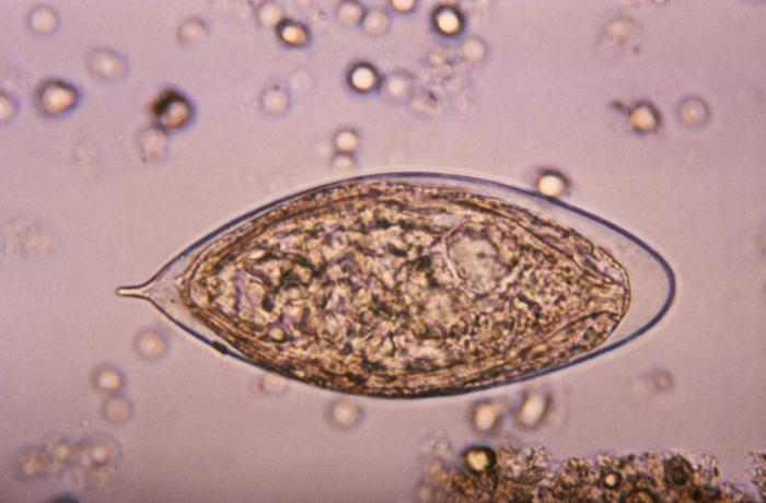
ID#:4843 This micrograph depicts an egg from a Schistosoma haematobium trematode parasite; magnified 500x. Note the egg's posteriorly-protruding, terminal spine, unlike the spinal remnant, which protrudes from the lateral wall of the Schistosoma japonicum egg. These eggs are eliminated in an infected human's feces or urine, and under optimal conditions in a watery environment, the eggs hatch and release "miracidia", which then penetrate a specific snail intermediate host. Once inside the host, the S. haematobium parasite passes through two developmental generations of sporocysts, and are released by the snail into its environment as "cercariae". |
|||
| 日期 | ||||
| 来源 | http://phil.cdc.gov/PHIL_Images/20031013/b47fc1793d7443d7a5cdbfbc73d95e53/4843_lores.jpg | |||
| 作者 | CDC, Public Health Image Library (PHIL) | |||
| 授权 (二次使用本文件) |
|
文件历史
点击某个日期/时间查看对应时刻的文件。
| 日期/时间 | 缩略图 | 大小 | 用户 | 备注 | |
|---|---|---|---|---|---|
| 当前 | 2006年5月7日 (日) 17:13 | | 700 × 460(38 KB) | Patho | {{Information| |Description=ID#: 4843 Description: This micrograph depicts an egg from the trematode parasite Schistosoma japonicum with its vestigial spine. The Schistosoma japonicum egg is typically oval or subspherical, has a vestigial spine, and is |
文件用途
全域文件用途
以下其他wiki使用此文件:
- ar.wikipedia.org上的用途
- cs.wikipedia.org上的用途
- de.wikibooks.org上的用途
- en.wikipedia.org上的用途
- fr.wikipedia.org上的用途
- gl.wikipedia.org上的用途
- ha.wikipedia.org上的用途
- nl.wikipedia.org上的用途
- sw.wikipedia.org上的用途
- tr.wikipedia.org上的用途